VolDiT
Controllable Volumetric Medical Image Synthesis
with Diffusion Transformers
MICCAI 2026
University Hospital Heidelberg Institute for Artificial Intelligence in Cardiovascular Medicine
High-resolution medical synthesis
Synthetic CT examples from LUNA16 and TaviCT.
Explore the full volume
Rotate, cut through, and inspect synthetic anatomy in three dimensions.
Loads on request · about 13 MB for the first volume and renderer.
Browser preview preserves the full field of view. Powered by NiiVue.
Anatomy, modeled in three dimensions
Native 3D tokens. Global context. Spatial control.
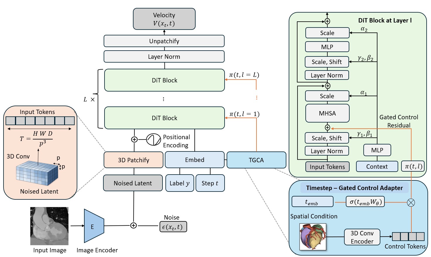
Compress the volume
A volumetric autoencoder maps a CT scan into a compact 3D latent space.
Learn global structure
A diffusion transformer models spatial relationships across native 3D tokens.
Guide the anatomy
A timestep-gated adapter adds segmentation guidance to a frozen backbone.
VolDiT combines compact volumetric representation with global self-attention. The same backbone supports unconditional generation and synthesis guided by segmentation masks.
See the evidenceGeneration quality, measured
Results from the MICCAI 2026 paper · 100 generated volumes per comparison.
| Model | FID ↓ | Precision ↑ | Recall ↑ |
|---|---|---|---|
| U-Net LDM | 0.031 | 0.44 | 0.94 |
| HA-GAN | 0.126 | 1.00 | 0.00 |
| VolDiT (L, p=4) | 0.004 | 0.91 | 0.90 |
| Model | FID ↓ | Precision ↑ | Recall ↑ |
|---|---|---|---|
| U-Net LDM | 36.8 | 0.95 | 0.63 |
| HA-GAN | 155.5 | 1.00 | 0.00 |
| VolDiT (L, p=2) | 21.4 | 0.94 | 0.73 |
FID uses different feature extractors; compare values only within each dataset.
Spatial control from segmentation masks
Median Dice on TaviCT · Learned timestep gate
Heart
- VolDiT
- 0.94
- U-Net
- 0.89
Aorta
- VolDiT
- 0.92
- U-Net
- 0.83
Fixed-size volumes and dataset-specific autoencoders. Synthetic examples illustrate generation, not clinical validity.
Full results and ablationsBuild on VolDiT
Paper, implementation, and directions for further research.
Research resources
Cite this work
@inproceedings{seyfarth2026volditcontrollablevolumetricmedical,
  title = {VolDiT: Controllable Volumetric Medical Image Synthesis with
           Diffusion Transformers},
  author = {Seyfarth, Marvin and Dar, Salman Ul Hassan and Frisch, Yannik
            and Wild, Philipp and Frey, Norbert and Andr{\'e}, Florian and
            Engelhardt, Sandy},
  booktitle = {Medical Image Computing and Computer Assisted Intervention --
               MICCAI 2026},
  year = {2027},
  publisher = {Springer Nature Switzerland},
  series = {Lecture Notes in Computer Science},
  volume = {16890},
  pages = {596--606},
  doi = {10.1007/978-3-032-38189-7_57}
}
Ongoing research VolDiT v2
Flow matching
Alternative objectives for training and sampling.
Spacing-aware positions
Position encodings that reflect volume geometry.
Latent normalization
Channel-wise scaling and controlled ablations.
These directions are under investigation; results above refer to the MICCAI 2026 paper.