CardioDiT
Latent Diffusion Transformers
for 4D Cardiac MRI Synthesis
MICCAI 2026
University Hospital Heidelberg Institute for Artificial Intelligence in Cardiovascular Medicine
A heart in four dimensions
Synthetic short-axis cine MRI · synchronized across depth
Explore cardiac motion
Inspect a synthetic 3D volume throughout the cardiac cycle.
Loads on request · all 32 frames of one synthetic sequence.
Coarse through-plane sampling; slice views retain the source detail. Powered by NiiVue.
Anatomy and motion, modeled together
Joint 3D+t synthesis in a compact latent space.
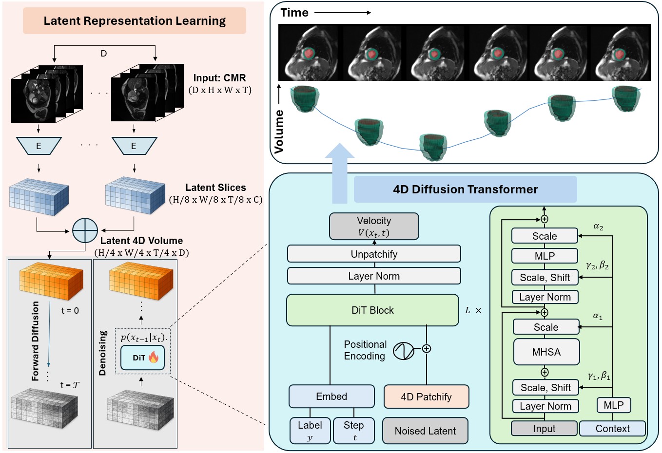
Encode the sequence
Compress each 2D+t slice with a spatiotemporal autoencoder.
Model space and time
Model stacked slice latents jointly with a 4D diffusion transformer.
Decode the motion
Decode the latent volume into a complete cardiac cine MRI sequence.
CardioDiT learns anatomy and cardiac motion jointly across a complete 3D+t volume. Four-dimensional patches preserve relationships across space and time.
See the evidenceGeneration quality, measured
Results from the MICCAI 2026 paper · 50 generated volumes per model.
| Model | FID ↓ | Precision ↑ | Recall ↑ |
|---|---|---|---|
| 2D+t LDM | 100.1 | 0.89 | 0.17 |
| 3D+t U-Net | 221.5 | 0.59 | 0.64 |
| CardioDiT | 71.9 | 0.97 | 0.70 |
| Model | FID ↓ | Precision ↑ | Recall ↑ |
|---|---|---|---|
| 2D+t LDM | 32.9 | 0.58 | 0.36 |
| 3D+t U-Net | 51.3 | 0.43 | 0.23 |
| CardioDiT | 21.2 | 0.82 | 0.54 |
Each cohort uses its real validation set as reference.
Cardiac function across the cycle
Ejection fraction · mean ± standard deviation (%)
Public cohorts
- CardioDiT
- 49.3 ± 15.1
- Real
- 47.6 ± 17.1
Private cohort
- CardioDiT
- 42.3 ± 6.3
- Real
- 44.6 ± 7.3
Fixed spatial sizes and 32-frame standardization. Full structural and function analyses are reported in the paper.
Full results and ablationsBeyond the original model
Ongoing experiments in spatial and temporal modeling.
Flexible geometry
Axis-aware 4D rotary position encodings and support for variable spatial shapes.
Native temporal lengths
Development toward sequences with variable frame counts.
Alternative objectives
Experiments with flow matching and self-conditioning.
The example is from a fixed-shape F07 run. These experiments are separate from the paper benchmarks; comparative gains are not yet established.
Resources & citationBuild on CardioDiT
Paper, implementation, and citation.
Research resources
- Read the paperMICCAI 2026 · Springer proceedings
- Explore the codeCardio-AI / cardiodit
- Download BibTeXCitation file
Pretrained weights are listed as forthcoming in the public repository.
Cite this work
@inproceedings{seyfarth2026cardiodit,
  title = {CardioDiT: Latent Diffusion Transformers for 4D Cardiac MRI
           Synthesis},
  author = {Seyfarth, Marvin and M{\"u}ller, Sarah Kaye and Ghanaat, Arman
            and Ayx, Isabelle and Fastenrath, Fabian and Wild, Philipp and
            Hertel, Alexander and Papavassiliu, Theano and Dar, Salman Ul
            Hassan and Engelhardt, Sandy},
  booktitle = {Medical Image Computing and Computer Assisted Intervention --
               MICCAI 2026},
  year = {2027},
  publisher = {Springer Nature Switzerland},
  series = {Lecture Notes in Computer Science},
  volume = {16890},
  pages = {69--79},
  doi = {10.1007/978-3-032-38189-7_7}
}